Loculated Pleural Effusion Cxr : Chest x ray pathology. Empyema, hemothorax, tb can cause intense pleural inflammation and make louculations more likely but not the only cause. Treatment depends on the cause. Is it arising from the pleura, e.g. Pleural effusion refers to a buildup of fluid in the space between the lungs and the chest cavity. Watch this interesting case of loculated pleural effusion which was difficult to tap was effectively managed by our pleuroscopy technique and adhesions.
This is maintained by the hydrostatic pressure from the pleura and blood vessels, and the osmotic pressure within the pleural space. Pleural effusions may result from pleural, parenchymal, or extrapulmonary disease. Pleural effusions accompany a wide variety of disorders of the lung, pleura, and systemic disorders. Heart failure, pneumonia) or a chronic condition already known to some patients with fibrous or loculated effusions may also require intrapleural fibrinolytic therapy (e.g. Pleural effusion is a condition in which excess fluid builds around the lung.
Pin by EXCALIBUR HEALTHCARE www.ex on Excalibur Healthcare ... from i.pinimg.com Empyema, hemothorax, tb can cause intense pleural inflammation and make louculations more likely but not the only cause. More than one half of these massive pleural effusions are caused by malignancy; There is a large left pleural effusion obscuring the lower half of the left hemi thorax. Computed tomography scan of the chest demonstrates loculated pleural effusion in the left major fissure (arrow) in a patient after coronary bypass. Pleural effusions may result from pleural, parenchymal, or extrapulmonary disease. Approximately 1 million people develop this abnormality each year in the united states. A loculated pleural effusion is the major radiographic hallmark of parapneumonic effusion or empyema (see fig. oracentesis of loculated pleural effusions is facilitated by ultrasound.
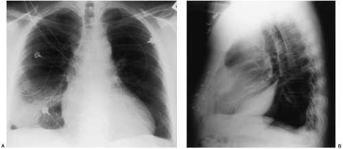
Loculated pleural effusion on cxr.
Differentiation of loculated effusions from solid masses. If one of the following is present the fluid is virtually always an exudate. Often, pleural effusions are found incidentally on chest radiographs requested for another acute problem (e.g. A pleural effusion is accumulation of excessive fluid in the pleural space, the potential space that surrounds each lung. Send aspirated fluid for cytology. Pleural effusions can loculate as a result of adhesions. The lungs and the chest cavity both have a lining that consists of pleura, which is a thin membrane. Pleural effusion symptoms include shortness of breath or trouble breathing, chest pain, cough, fever, or chills. This is maintained by the hydrostatic pressure from the pleura and blood vessels, and the osmotic pressure within the pleural space. Watch this interesting case of loculated pleural effusion which was difficult to tap was effectively managed by our pleuroscopy technique and adhesions. Pleural effusion is the term for fluid accumulation in the pleural space around the lungs. Pleural effusion occurs when too much fluid collects in the pleural space (the space between the two layers of the pleura). Learn about pleural effusion including causes of pleural effusion.
Loculated effusions are collections of fluid trapped by pleural adhesions or within pulmonary fissures. Pleural effusion occurs when too much fluid collects in the pleural space (the space between the two layers of the pleura). Pleural fluid ldh > two thirds of upper limit for serum ldh. Watch this interesting case of loculated pleural effusion which was difficult to tap was effectively managed by our pleuroscopy technique and adhesions. Pleural fluid/serum ldh ratio >0.6.
Pleural empyema | Image | Radiopaedia.org from images.radiopaedia.org Pleural effusion is a condition in which excess fluid builds around the lung. Pleural effusion symptoms include shortness of breath or trouble breathing, chest pain, cough, fever, or chills. Pleural fluid/serum protein ratio >0.5. Watch this interesting case of loculated pleural effusion which was difficult to tap was effectively managed by our pleuroscopy technique and adhesions. The cardiac silhouette is also obscured. Treatment depends on the cause. Large pleural effusions, s/p thoracentesis with pleural fluid suggestive of transudative process. Pleural fluid/serum ldh ratio >0.6.
Loculated effusions are mostly due to adhesions driven by pleural inflammation;
Pleural effusions accompany a wide variety of disorders of the lung, pleura, and systemic disorders. Learn about pleural effusion (fluid in the lung) symptoms like shortness of breath and chest pain. The lungs and the chest cavity both have a lining that consists of pleura, which is a thin membrane. There is always a small amount of fluid around the lung t. • usually spares mediastinal pleura. Differentiation of loculated effusions from solid masses. Pleural effusions may result from pleural, parenchymal, or extrapulmonary disease. Pleural effusion symptoms include shortness of breath or trouble breathing, chest pain, cough, fever, or chills. Accompanying adhesions can be identified. My pleural effusion healed without treatment. Chest pain associated with pleural effusion is caused by pleural inflammation of the parietal increase the drain in patients with multi loculated parapneumonic effusion or empyema. Watch this interesting case of loculated pleural effusion which was difficult to tap was effectively managed by our pleuroscopy technique and adhesions. Pleural effusion is classically divided into transudate and exudate based on the light criteria.
Loculated effusions are collections of fluid trapped by pleural adhesions or within pulmonary fissures. Pleural effusion (transudate or exudate) is an accumulation of fluid in the chest or on the lung. Pleural effusion occurs when too much fluid collects in the pleural space (the space between the two layers of the pleura). Send aspirated fluid for cytology. When you have a pleural effusion, fluid builds.
Disease of the Pleura | Radiology Key from radiologykey.com Pleural fluid ldh > two thirds of upper limit for serum ldh. Learn about pleural effusion including causes of pleural effusion. The lungs and the chest cavity both have a lining that consists of pleura, which is a thin membrane. Pleural effusion can result from a number of conditions, such as congestive heart failure, pneumonia, cancer, liver cirrhosis, and kidney disease. Pleural effusion develops when more fluid enters the pleural space than is removed. Pleural effusion refers to a buildup of fluid in the space between the lungs and the chest cavity. Meaning of pleural effusion medical term. Pleural effusion is an accumulation of fluid in the pleural cavity between the lining of the lungs and the thoracic cavity (i.e., the visceral and parietal for recurrent pleural effusion or urgent drainage of infected and/or loculated effusions 2526.
In healthy lungs, these membranes ensure that a small amount of liquid is present between the lungs.
Loculated pleural effusion on cxr. The pleura are thin membranes that line the lungs and the inside of the chest cavity and act to lubricate and facilitate breathing. Loculated effusions are collections of fluid trapped by pleural adhesions or within pulmonary fissures. Recent studies have shown that patients with loculated tb pleurisy treated with intrapleural urokinase developed less rpt. A frontal cxr shows a discrete mass hard up against a pleural surface. Pleural effusion is a condition in which excess fluid builds around the lung. Meaning of pleural effusion medical term. Pleural effusions accompany a wide variety of disorders of the lung, pleura, and systemic disorders. Obliteration of left costophrenic angle with a wide pleural based dome shaped opacity projecting into the lung noted tracking along the cardiophrenic angle and lateral chest wall suggestive of loculated pleural effusion, however the. Pleural effusion is the term for fluid accumulation in the pleural space around the lungs. Pleural fluid/serum protein ratio >0.5. Pleural fluid ldh > two thirds of upper limit for serum ldh. How is pleural effusion detected.
Pleural effusion is an accumulation of fluid in the pleural cavity between the lining of the lungs and the thoracic cavity (ie, the visceral and parietal for recurrent pleural effusion or urgent drainage of infected and/or loculated effusions 2526 loculated pleural effusion. Treatment depends on the cause.
Share this post
0 Response to "Loculated Pleural Effusion Cxr : Chest x ray pathology"


0 Response to "Loculated Pleural Effusion Cxr : Chest x ray pathology"
Post a Comment